
[box type=”bio”] Learning Point of the Article: [/box]
In addition to proper patient selection and surgical technique, computer-assisted navigation serves as a reliable tool to improve component alignment and clinical outcomes after hip resurfacing.
Case Report | Volume 9 | Issue 3 | JOCR May-June 2019 | Page 93-97 | Ameer Elbuluk, Karlina Fiaes, Jessica R. Benson, Edwin Su. DOI: 10.13107/jocr.2250-0685.1440
Authors: Ameer Elbuluk[1], Karlina Fiaes[2], Jessica R. Benson[3], Edwin Su[1]
[1]Department of Orthopaedics, Hospital for Special Surgery, New York, NY 10021, United States.
[2]Department of Health Studies, Faculty of Applied Health Sciences, School of Public Health and Health Systems University of Waterloo, 200 University Avenue West, Waterloo, Ontario N2L 3G1, Canada.
[3]Department of Clinical Research, Intellijoint Surgical, Inc., 809 Wellington Road North, Kitchener, ON, N2H 5L6, Canada.
Address of Correspondence:
Dr. Ameer Elbuluk,
Department of Orthopaedics, Hospital for Special Surgery, New York, NY 10021, United States.
E-mail: elbulukam@hss.edu
Abstract
Introduction: The ReCap Femoral Resurfacing System has been associated with increased cases of revision surgery when compared to other hip resurfacing systems. However, computer-assisted navigation may have the potential to reduce the risk of post-operative complications by providing more accurate intraoperative measurements for acetabular component positioning.
Case Report: The present case describes an active 46-year-old male presenting with severe osteoarthritis of the right hip who elected to undergo a ReCap resurfacing arthroplasty with navigation. Results demonstrated accurate acetabular component position and leg length measurements to within <1° and 1mm of standard radiographic measurements.
Conclusion: These findings are the first to describe the use of navigation with the ReCap system and provide encouraging results for further clinical evaluation.
Keywords: Accuracy, Computer-assisted navigation, Cup position, Hip resurfacing arthroplasty, Leg length, ReCap Femoral Resurfacing System hip resurfacing arthroplasty; Computer-assisted navigation; ReCap Femoral Resurfacing System; Cup Position; Leg Length; Accuracy
Introduction
Common advantages of hip resurfacing arthroplasty have been cited as conservation of femoral bone, quick return to physical activity, and a reduced dislocation rate[1, 2, 3]. In particular, the ReCap® Femoral Resurfacing System (Biomet Inc., Warsaw, IN) has demonstrated excellent survivorship [4], but reports indicate a larger risk of revision surgery in comparison to the more commonly used Birmingham Hip Resurfacing System (BHR; Smith and Nephew, Andover, MA) [5, 6]. In part, hip resurfacing revision rate has been attributed to dislocation and malalignment [5, 6], which underscores the importance of proper component positioning. Indeed, malposition is associated with increased component loosening, impingement, edge loading and wear, and early dislocation [4, 7, 8]. Computer-assisted navigation systems (CASs) are an emerging technology that can assist with component placement intraoperatively. The value of CAS in resurfacing arthroplasty has been previously demonstrated, with studies reporting accurate femoral and acetabular component placement [9, 10, 11, 12], and a potentially reduced learning curve when compared to conventional surgical techniques [10]. However, to the best of our knowledge, the utilization of CAS with the ReCap Femoral Resurfacing System has never been described. The present report summarizes a case of resurfacing arthroplasty utilizing the ReCap Femoral Resurfacing System, in which CAS was used to assist with accurate intraoperative acetabular component positioning.
Case Report
Patient information
A 46-year-old male presented with a chief complaint of progressively worsening right hip pain over the past decade. The patient denied antecedent trauma or any hip injuries since childhood. The patient did note that he was previously very active and went running and cycling on a regular basis. However, he became limited due to pain in his groin, buttock, and thigh, which were all exacerbated by weight-bearing activities. On a day-to-day basis, the patient reported difficulty with stairs, limping while ambulating, and significant trouble getting in and out of a car. The patient’s medical history was significant for a lumbar disc herniation 8 years prior, for which he underwent a microdiscectomy at L5-S1. Past medical, family and social history were otherwise unremarkable. Conservative management, including nonsteroidal anti-inflammatory medications, activity modification, and physical therapy, only provided minimal relief.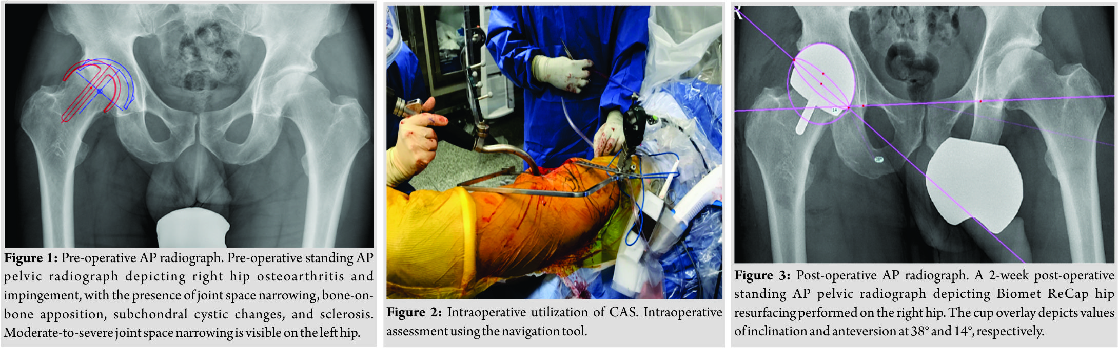
Diagnostic assessment
Initial orthopedic examination revealed the patient walking with a coxalgic gait and abductor lurch to the right. To test for anterior impingement, while lying supine, the patient’s hip was internally rotated and adducted during passive flexion to 90°, which reproduced the patient’s symptom of groin pain. The hip was able to internally rotate to neutral and externally rotate to 30°. On range of motion testing, abduction was 30°and the patient had 0°of adduction. Patrick’s test (flexion, abduction, and external rotation) was also positive and associated with severe groin pain and restricted sacroiliac joints. There were groin and buttock pain with passive hip motion in all directions. Lower limb neurologic and vascular examination was unremarkable. Plain film radiographs revealed severe right hip osteoarthritis with bone-on-bone apposition, subchondral cysts and sclerosis (Fig. 1). A rounded focus of ossification adjacent to the acetabulum was found, which may have reflected os acetabuli or ossified labrum. Cam-type configurations were seen bilaterally. The left hip also demonstrated moderate-to-severe joint space narrowing. Degenerative changes of bilateral sacroiliac joints were also identified. A lengthy discussion was had regarding hip resurfacing versus hip replacement; however, based on the patient’s age and activity level, conservative hip resurfacing with metal-on-metal (MoM) parts was deemed an appropriate option. The patient agreed, given his desire to run at least 5 miles twice per week, competed in triathlons, and work. Risks and benefits were discussed in detail with the patient. We discussed surgical approaches to the hip joint and the rationale for a posterior approach.
Treatment
The patient was brought to the operating room and was placed on the hip table in the lateral decubitus position. The patient’s surgical area was prepped and draped in the standard, sterile fashion. Two threaded pins for a minioptical navigation tool (Intellijoint HIP®, Intellijoint Surgical Inc., Waterloo, ON, Canada; off-label use) were placed into the iliac crest, approximately 2 cm posterior to the ASIS. The navigation system camera was then attached, and registration was performed. Next, a standard posterolateral incision was made through the skin and dissection was carried down through subcutaneous tissue to the underlying fascia achieving hemostasis where necessary. The posterior short external rotators were identified, and the piriformis, conjoined tendons, and quadratus femoris were detached and tagged. Following elevation of the gluteus minimus, a 360° circumferential capsulotomy was performed. The femoral disc for navigational leg length measurements was impacted onto the greater trochanter, and registration of the hip center of rotation was performed. Once the hip was dislocated, the femoral head and neck were exposed. Osteophytes from the anterior superior aspect of the femoral neck were now removed, restoring the anterior head-neck offset. The femoral head was then measured as to its diameter. The head-neck templates were placed along the posterior aspect of the femoral neck to measure the sizing. Lines were marked along the midpoint of the femoral neck in both the coronal and sagittal planes. At this point, attention was turned toward the acetabulum. The labrum, as well as the tissues from the acetabular fossa, was removed. Reaming was begun, with the plan for a 1 mm press-fit. Reaming was first directed medially and then in the desired alignment of the acetabular implant. A trial implant was impacted into place and marked as to its depth of insertion using electrocautery. The actual implant (58mm M2a-Magnum cup; Biomet Inc., Warsaw, IN, USA) was then placed on its insertion handle and impacted into place. The alignment was checked using external alignment guides, bony landmarks, and corroboration with pre-operative templating. The navigation unit was then used to confirm final acetabular component position, measuring 14° of anteversion and 39° of inclination as selected by the surgeon intraoperatively. Following femoral preparation, the trial femoral implant was passed around the prepared bone to ensure adequate bone preparation and contact. With the trial femoral implant in place, the hip was reduced and the navigation device used to confirm the restoration of leg lengths, measuring 4 mm of lengthening (Fig. 2). The hip was again dislocated and the trial head removed. It was then decided that the bone was sufficiently supportive to allow for uncemented fixation and the actual implant was impacted. The head was relocated into the acetabulum, after ensuring that there was no debris or soft tissue interposed. The capsule and short external rotators were reattached through a bony bridge in the greater trochanter. The quadratus femoris and gluteus maximus tendon insertion were also repaired. The navigation hardware was then removed. We returned the patient to the supine position. We verified that all lower extremity compartments were soft and compressible and that we had intact distal pulses. The patient was then transferred to the recovery room in stable condition.
Post-operative outcomes
Standard, pre- and post-operative AP pelvic radiographs were obtained and analyzed using TraumaCad (Brainlab, Chicago, USA). Final values for cup position and leg length were measured in triplicate and averaged. Radiographic analysis revealed a final post-operative cup position of 14.3° anteversion and 38.3° inclination on the post-operative radiograph (Fig. 3), as well as a lengthened operative leg of 5 mm between pre- and post-operative images.
Follow-up
At 1-year post-operative, the patient was doing extremely well, demonstrating range of motion in the operative hip of 0°–120° flexion, 40° external rotation, 10° internal rotation, 40° abduction, and 10° adduction. Harris Hip Score was 91.8, with no instability in the joint and both neuromuscular and vascular examinations normal. The patient was able to walk unlimited distances, run, cycle and swim with no physical limitations.
Discussion
Hip resurfacing arthroplasty was first popularized in the 1960s but was largely limited by high rates of component wear, loosening, and failure attributed to poor materials and design [1, 13]. Decades later, technological advancements combined with MoM prostheses helped resurfacing arthroplasty regain popularity, with improved long-term survivorship results up to 7 years postoperatively [5, 6]. While surgical benefits include conserved femoral bone stock and quick return to an active lifestyle, acetabular component mal positioning can result in adverse outcomes such as edge loading, accelerated wear, impingement, instability, and loosening [4, 7, 8]. Revision surgery following resurfacing arthroplasty has been associated with malpositioned components, and the ReCap Femoral Resurfacing System, in particular, has been reported with a higher likelihood of revision than the more commonly used BHR [5, 6]. CAS has demonstrated accurate measurements of cup position during cases of BHR [10, 11, 12]and may help to reduce revision rates by improving component placement during surgery. However, utilization of CAS with the ReCap system specifically has yet to be addressed. Given these criteria, the current case report sought to examine whether computer-assisted navigation during a ReCap resurfacing procedure could provide intraoperative measurements of acetabular component position with accuracy. Recent results from national joint registries have reported revision rates for the ReCap system to range between 3.4% and 8.7% at 3 years postoperatively, and between 7.8% and 12.2% at 7 years [5, 6]. In comparison, a reduced likelihood of revision for BHR was reported as 2.4% at 3 years and between 4.9% and 5.5% at 7 years. While the probability of revision surgery is higher for the ReCap system in comparison to the commonly used BHR system, a report by Gross and Liu [4] described an improved 7-year revision rate in a series of hybrid ReCap resurfacing procedures utilizing the Biomet M2a-Magnum acetabular component. Specifically, the study reported 96.7% survivorship rate for the ReCap-Magnum system (3.4% revision rate), which demonstrated an improvement in revision rate from a previously reported 5.7% [14]. Interestingly, the improved survivorship rate observed is congruent with those reported for other resurfacing systems [15, 16, 17, 18]. Correct component positioning is imperative to reducing the risk of post-operative complications that may lead to revision surgery. The present case demonstrated the ability of CAS to accurately measure cup position and leg length in a resurfacing case utilizing the hybrid ReCap-Magnum system described above. Intraoperative navigational measurements for cup position were recorded at 14° of anteversion and 39° of inclination, confirmed on post-operative radiographic analysis which revealed a final cup position of 14.3° anteversion and 38.3° inclination. Intraoperative leg length was also confirmed by the navigational tool, recording 4 mm of lengthening following final implant placement. This corresponded postoperatively with a calculated radiographic leg length measurement of 5 mm. Effectively, a pre-operative leg length differential (LLD) of 6 mm was restored to 1 mm postoperatively, and the device accurately monitored leg length throughout the procedure. The results observed in this novel report evaluating CAS utilization with the ReCap resurfacing system are consistent with several previous findings in hip resurfacing. For acetabular component positioning, the navigation tool used in the present report has previously demonstrated accuracy to within 0.7°–3° of standard radiographic measurements in cases of BHR [11, 12]. The navigation tool used in this case can also effectively monitor and restore leg length intraoperatively [12] and has demonstrated the ability to account for and monitor cases with significant LLD, as demonstrated in a previous case of complex Legg-Calve-Perthes disease [19]. The present case reported acetabular component positioning accuracy between 0.3° and 0.7°, exhibiting promising results for the use of CAS in resurfacing arthroplasty performed with the ReCap system. Additionally, intraoperative leg length monitoring effectively predicted LLD restoration following the procedure. Limitations of the present work include the inability of the navigation tool to assist with femoral pin placement; however, this was accounted for with thorough pre-operative planning and intraoperative templating. The device was able to monitor leg length changes throughout surgery and accurately recorded leg length to within 1 mm of standard measurements. Second, this report provides a singular case regarding CAS utilization with the ReCap system. The veracity with which report conclusions can be broadly applied is minimal. However, the findings in this report present encouraging results for the integration of CAS with the ReCap system and support further clinical evaluation moving forward.
Conclusion
The current case report demonstrates accurate results utilizing novel CAS in resurfacing arthroplasty performed with the ReCap Femoral Resurfacing System. The navigational tool exhibits accurate acetabular component placement and the ability to monitor changes in leg length. This report adds favorable evidence to a growing database of navigation research in resurfacing arthroplasty; however, further clinical evidence is still required. CAS may be a beneficial tool in the armamentarium of hip surgeons performing resurfacing procedures.
Clinical Message
Computer-assisted navigation may assist with the accuracy of acetabular component positioning during ReCap resurfacing arthroplasty.
References
1. Sershon R, Balkissoon R, Valle CJ. Current indications for hip resurfacing arthroplasty in 2016. Curr Rev Musculoskelet Med 2016;9:84-92.
2. Jameson SS, Baker PN, Mason J, Porter ML, Deehan DJ, Reed MR, et al. Independent predictors of revision following metal-on-metal hip resurfacing: A retrospective cohort study using national joint registry data. J Bone Joint Surg Br 2012;94:746-54.
3. American Joint Replacement Registry. Fourth AJRR Annual Report on Hip and Knee Arthroplasty; 2017.
4. Gross TP, Liu F. Hip resurfacing with the biomet hybrid reCap-magnum system: 7-year results. J Arthroplasty 2012;27:1683-900.
5. National Joint Registry. 13th Annual Report; 2016.
6. Australian Orthopaedic Association. Hip and Knee Arthroplasty Annual Report; 2015.
7. Campbell P, Beaulé PE, Ebramzadeh E, Le Duff MJ, De Smet K, Lu Z, et al. The john charnley award: A study of implant failure in metal-on-metal surface arthroplasties. Clin Orthop Relat Res 2006;453:35-46.
8. De Haan R, Campbell PA, Su EP, De Smet KA. Revision of metal-on-metal resurfacing arthroplasty of the hip: The influence of malpositioning of the components. J Bone Joint Surg Br 2008;90:1158-63.
9. El Hachmi M, Penasse M. Our midterm results of the birmingham hip resurfacing with and without navigation. J Arthroplasty 2014;29:808-12.
10. Romanowski JR, Swank ML. Imageless navigation in hip resurfacing: Avoiding component mal position during the surgeon learning curve. J Bone Joint Surg Am 2008;90 Suppl 3:65-70.
11. Vigdorchik JM, Elbuluk A, Benson JR, Muir JM. Birmingham hip resurfacing using a novel mini-navigation system: A Case report. J Orthop Case Rep 2018;8:48-52.
12. Shah R, Benson JR, Muir JM. Computer-assisted navigation in birmingham hip resurfacing: A case report. SAGE Open Med Case Rep 2018;6:1-5.
13. Grigoris P, Roberts P, Panousis K, Jin Z. Hip resurfacing arthroplasty: The evolution of contemporary designs. Proc Inst Mech Eng H 2006;220:95-105.
14. van der Weegen W, Hoekstra HJ, Sijbesma T, Austen S, Poolman RW. Hip resurfacing in a district general hospital: 6-year clinical results using the reCap hip resurfacing system. BMC Musculoskelet Disord 2012;13:247.
15. Lilikakis AK, Vowler SL, Villar RN. Hydroxyapatite-coated femoral implant in metal-on-metal resurfacing hip arthroplasty: Minimum of two years follow-up. Orthop Clin North Am 2005;36:215-22, 9.
16. McBryde CW, Theivendran K, Thomas AM, Treacy RB, Pynsent PB. The influence of head size and sex on the outcome of birmingham hip resurfacing. J Bone Joint Surg Am 2010;92:105-12.
17. Treacy RB, McBryde CW, Pynsent PB. Birmingham hip resurfacing arthroplasty. A minimum follow-up of five years. J Bone Joint Surg Br 2005;87:167-70.
18. Amstutz HC, Le Duff MJ. Eleven years of experience with metal-on-metal hybrid hip resurfacing: A review of 1000 conserve plus. J Arthroplasty 2008;23:36-43.
19. Shah RR, Gobin V, Muir JM. Imageless navigation improves intraoperative monitoring of leg length changes during total hip arthroplasty for legg-calve-perthes disease: Two case reports. Case Rep Orthop 2018;2018:4362367.
 |
 |
 |
 |
| Dr. Ameer Elbuluk | Ms. Karlina Fiaes | Ms. Jessica R. Benson | Dr. Edwin Su |
| How to Cite This Article: Elbuluk A, Fiaes K, Benson JR, Su E. Computer-assisted Navigation in Hip Resurfacing Arthroplasty: A Case Study utilizing the ReCap Femoral Resurfacing System. Journal of Orthopaedic Case Reports 2019 May-June; 9(3): 93-97. |
[Full Text HTML] [Full Text PDF] [XML]
[rate_this_page]
Dear Reader, We are very excited about New Features in JOCR. Please do let us know what you think by Clicking on the Sliding “Feedback Form” button on the <<< left of the page or sending a mail to us at editor.jocr@gmail.com


