[box type=”bio”] What to Learn from this Article?[/box]
Unusual Presentation of Tumoral Calcinosis.
Case Report | Volume 4 | Issue 3 | JOCR July-Sep 2014 | Page 59-62 |Banshelkikar SN, Argekar H, Bhoir A. DOI: 10.13107/jocr.2250-0685.199
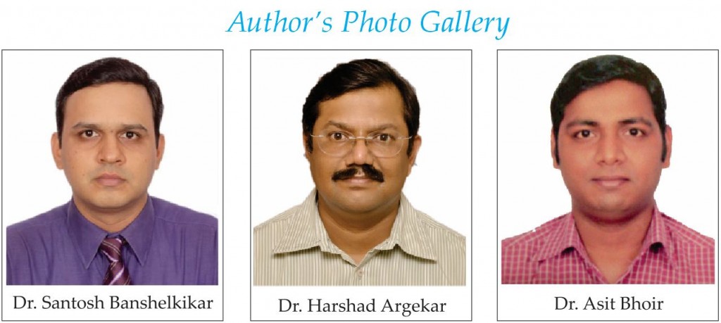
Authors: Banshelkikar SN[1], Argekar H[1], Bhoir A[1]
[1] Department Of Orthopaedics, Lokmanya Tilak Municipal Medical College And General Hospital, Sion. Mumbai. India.
Address of Correspondence:
Dr. Santosh N Banshelkikar, Flat No 101, Bldg No 40/1, Siddhivinayak Chs, Near Municipal School, Tilaknagar – West. Mumbai – 400089. Mobile: +91 9930 40 21 22, Email: drsantoshnb@gmail.com
Abstract
Introduction: Tumoral calcinosis is an uncommon disorder characterised by the deposition of calcium phosphate in periarticular tissues. The deposits are usually around large joints; but rarely can be found around small joints of hand and feet.
Case Report: We present the case of 13 year old female with three years history of spontaneous, progressively increasing, painful swellings along right middle finger and right heel. She was otherwise well and had normal serum calcium but elevated phosphate levels. Plain radiography demonstrated a dense lobulated cluster of calcific nodules within soft tissues consistent with a diagnosis of tumoral calcinosis. This diagnosis was confirmed on the basis of histopathological examination following surgical excision.
Conclusion: As such tumoral calcinosis is a rare entity and with such unusual presentations like in our case, it may lead to diagnostic confusion. Tumoral calcinosis should be considered in the differential diagnosis of painful swellings developing in the vicinity of small joints of hand and feet.
Keywords: Tumoral calcinosis , small joints , calcium salts deposition.
Introduction
The first report of tumoral calcinosis was by duret [1] in 1899 who termed it “Endothelium calcifie”. The term tumoral calcinosis was proposed by inclan [2] in 1943 and was accepted worldwide. Tumoral calcinosis is an uncommon pathological entity characterised by multiple circumscribed calcified masses in periarticular connective tissue. The name indicates calcinosis (calcium deposition) which resembles like tumour; but they are not true neoplasms as they do not have dividing cells. These lesions mainly comprise calcium hydroxy apatite crystals and amorphous calcium phosphate [3]. They are commonly located around large joints like hip, elbow, and shoulder with predilection for extensor surfaces. Adolescents and young adults are commonly affected. The usual presentation is firm, rubbery masses which are painless and non-tender [4]. The term tumoral calcinosis has also been loosely used to describe secondary metastatic peri articular calcification occurring in conditions such as hyperparathyroidism, renal insufficiency, hypervitaminosis D. Hyperphosphatemic familial tumoral calcinosis (HFTC) is an autosomal recessive entity with higher incidence in patients of African descent, characterised by an increase in the levels of phosphate in the blood and abnormal depositionof phosphate and calcium in body tissues [5]. The differential diagnosis includes calcium pyrophospha[e dehydrate deposition disease, soft tissue chondromas, myositis ossificans, dystrophic calcifications [6].
Case Report
A thirteen year old ,right hand dominant female presented to us following three years history of multiple , slowly growing masses on her right middle finger and right heel with scars of previous surgical excision at medial aspect of left elbow, right forearm extensor aspect and left foot lateral border. Mass lesions were painful and interfered with activities of daily routine. Her past medical as well as family history was not relevant. Histopathology report of previously excised swellings gave impression of epidermal keratinous cyst with calcific debris. The clinical examination revealed- I) A solitary 1.5x3cm elliptical lesion over volar aspect of middle phalanx of right middle finger. ii) Thickened area of 2×3 cm with ill defined margins over right heel. These swellings had a firm nonfluctuant nodular consistency with an overlying centrally located punctum from which no discharge was expressible. She had no regional lymphadenopathy. The range of motion at adjacent interphalangeal joints was full and free. Radiograph showed multiple round to oval calcified masses along volar surface of right middle finger and plantar surface of right heel. Her laboratory investigations like blood counts, erythrocyte sedimentation rate, serum uric acid, serum calcium, alkaline phosphate were within normal range; only serum phosphate levels were slightly raised. Surgical excision was performed through volar approach for finger mass and through plantar approach for heel lesion. The excised material consisted of lobulated yellowish masses with well defined capsules. Cut surfaces of the lobules were yellowish white with chalky granular deposits. Microscopically sections revealed thin and coarse calcific deposits with foreign body type of giant cells, few plasma cells and lymphocytes, surrounded by fibrosis. These features were compatible with tumoral calcinosis. Post operative radiographs confirmed the complete removal of the calcified tissue. She had no recurrence at her follow up visits. (mri and bone scan were not done, excision biopsy was preferred).
Discussion
Most of the literature describes painless nature of the swellings in tumoral calcinosis, mostly located around large joints of the body. The involvement of small joints of hand and feet is considered rare. Harkess and peters [4] reported 33 cases of tumoral calcinosis, out of which only one had hand involvement. In 2006 kamath et al [7] reported a case of tumoral calcinosis involving 2nd metacarpo phalangeal joint. In 2006 kim et al [8] reported 3 cases with metacarpo phalangeal and proximal interphalangeal joint involvement. Murai et al [9] reported a case of bilateral index finger involvement in a 5 year old male. Tumour calcinosis is usually seen on the extensor surfaces of large joints hip, shoulder, elbow in adolescents and young adults. These swellings are usually painless, slowly growing, and progressive in nature [4]. Blood chemistry shows normal calcium level and hyperphosphatemia . Standard radiographs show varying sized irregular shaped calcific deposits in peri articular soft tissue [3]. Our case had two distinctive features compared to most of the cases described in literature I) Calcific deposits were seen around small joints of hand like proximal and distal interphalangeal joints ii) Hand and heel lesions were painful in contrast to painless nature of the swellings at other body locations described in literature Painful nature of these swellings can possibly be due to superficial location, prone to friction and pressure on adjacent cutaneous nerves [10, 12]. Exact etiology is still unknown. However few pathologic mechanisms are implicated I) Mutations in genes coding for FGF23 or proteins controlling their activities (GALNT3 and KL gene) are involved [5, 11] ii) Local trauma has also been implicated. In 2011 nick hutt et al reported a case with post traumatic acral tumour calcinosis [12] In our case probably juxta articular injury has lead to reparative response with resultant synovial metaplasia forming bursa with deposition of calcium-phosphate product . [11] Other conditions with calcific deposits should be considered in Differential diagnosis like dystrophic calcifications, metastatic calcifications, heterotopic ossifications, soft tissue sarcomas, myositis ossificans, autoimmune diseases like dermatomyositis, scleroderma. [6, 13, 14, 15] Surgical excision is the mainstay of treatment of tumoral calcinosis with low recurrence rate. Phosphate depletion therapy for hyperphosphatemia can also be tried as adjuvant therapy.
Conclusion
Tumour calcinosis generally occurs around large joints; involvement of small joints like hand and feet as in our case,is extremely rare. We conclude that tumoral calcinosis should be considered in the differential diagnosis of painful swellings developing in the vicinity of small joints of hand and feet.
References
1. Duret M. Tumeurs multiples et singulaires desbourses sereuses. Bull Mem Soc Anthropal Paris 1899;74:725-32.
2. Inclan A. Tumoral calcinosis. JAMA 1943;121:490-5.
3. Grainger RG, Allison D, Adam A, Dixon AK. Diagnostic radiology: a textbook of medical imaging. 4th Ed; Churchill Livingstone, Philadelphia, 2001: Vol -3, p – 2085. 49:721-731,1967.
4. Harkess JW, Peters HJ. Tumoral calcinosis. A report of six cases. J Bone Joint Surg Am. 1967; 49(4):721-731. (http://jbjs.org/article.aspx?articleid=14711 )
5. Topaz O, Shurman DL, Bergman R. Mutations in GALNT3, encoding a protein involved in O-linked glycosylation, cause familial tumoral calcinosis. Nat Genet. 2004; 36(6):579-581.
6. University of Washington; dept of radiology, musculoskeletal radiology (http://www.rad.washington.edu/academics/academic -sections/msk/teaching-materials/online- musculoskeletal-radiology-book/ soft-tissue-calcifications).
7. Kamath BJ, Pinto D, Sharma C. Tumoral calcinosis of hand: a rare location with unusual presentation. Int J Orthop Surg. 2006; 3(3):22.
8. Kim HS, Suh JS, Kim YH, Park SH. Tumoral calcinosis of the hand: three unusual cases with painful swelling of the small joints. Arch Pathol Lab Med 2006;130:548–51 (http://www.ncbi.nlm.nih.gov/pubmed/16594750)
9. Murai S, Matsui M, Nakamura A. Tumoral calcinosis in both index fingers: a case report. Scand J Plast Reconstr Surg Hand Surg 2001;35:433–5. doi:10.1080/028443101317149426 (http://www.ncbi.nlm.nih.gov/pubmed/11878182 ).
10. Ozcelik c Aydogdu S, Doganavsargil B, Sur H:Tumoral calcinosis of the hand.Orthopedics2008,31:1145 (http://www.healio.com/orthopedics/hand-wrist/journals/ortho/%7B87d99b1a-489b-4689-b292-d2526eaeaf5d%7D/tumoral-calcinosis-of-the-hand )
11. Slavin RE, Wen J, Barmada A.Tumoral calcinosis–a pathogenetic overview: a histological and ultrastructural study with a report of two new cases, one in infancy. Int J Surg Pathol. 2012 Oct;20(5):462-73. Epub 2012 May 21
12. Nick Hutt, Davinder PS Baghla, Vivek Gulati Acral post-traumatic tumoral calcinosis in pregnancy: a case report; Journal of Medical Case Reports2011,5:89
http://www.jmedicalcasereports.com/content/5/1/89 JOURNAL OF MEDICALCASE REPORTS
(http://www.jmedicalcasereports.com/content/5/1/89 )
13. Resnick D, Niwayama G Clinical, radiographic and pathologic abnormalities in calcium pyrophosphate dihydrate deposition disease (CPPD): pseudogout. Radiology. 1977 Jan;122(1):1-15.
(http://www.ncbi.nlm.nih.gov/pubmed/186841).
14. Alexis Lacout, Mohamed Jarraya ;Myositis ossificans imaging: keys to successful diagnosis ; Indian journal of radiology and imaging ; 2013 jan –march 22(1) 35-39
(http://www.ncbi.nlm.nih.gov/pmc/articles/PMC3354355/).
15. Clayton E. Wheeler; Arthur c ;soft tissue calcification, with special reference to its occurrence in collagen diseases; annals of internal medicine; april 1952
(http://annals.org/article.aspx?articleid=674946).
16. Alkhooly AZ ; Medical treatment for tumoral calcinosis with eight years of follow-up: a report of four cases. J Orthop Surg (Hong Kong). 2009 Dec;17(3):379-82.
(http://www.ncbi.nlm.nih.gov/pubmed/20065385?dopt=Abstract).
| How to Cite This Article: Banshelkikar SN, Argekar H, Bhoir A. Idiopathic Tumoral Calcinosis with Unusual Presentation- Case Report with Review of Literature. Journal of Orthopaedic Case Reports 2014 July-Sep;4(3): 59-62. Available from: https://www.jocr.co.in/wp/2014/07/11/2250-0685-199-fulltext/ |
(Figure 1)|(Figure 2)|(Figure 3)|(Figure 4)|(Figure 5)|(Figure 6)|(Figure 7)|(Figure 8)|(Figure 9)|(Figure 10)
[Abstract] [Full Text HTML] [Full Text PDF] [XML]
[rate_this_page]
Dear Reader, We are very excited about New Features in JOCR. Please do let us know what you think by Clicking on the Sliding “Feedback Form” button on the <<< left of the page or sending a mail to us at editor.jocr@gmail.com


