
[box type=”bio”] What to Learn from this Article?[/box]
Mucoid degeneration of ACL and PCL is ussualy asymptomatic, but may present with various symptoms and artial debulking is beneficial.
Case Report | Volume 5 | Issue 4 | JOCR Oct-Dec 2015 | Page 44-46| Joon Ho Wang, Rajat R Jangir. DOI: 10.13107/jocr.2250-0685.343 .
Authors: Joon Ho Wang[1], Rajat R Jangir[1]
[1] Department of Orthopaedic Surgery, Samsung Medical Center, Sungkyunkwan University, School of Medicine, #81, Irwon-Ro, Gangnam-gu, Seoul, 135-710, Korea.
Address of Correspondence
Dr. Rajat R Jangir,
Department of Orthopaedic Surgery, Samsung Medical Center, Sungkyunkwan University, School of Medicine, #81, Irwon-Ro, Gangnam-gu, Seoul, 135-710, Korea. E mail – dr.rajatjangir@gmail.com
Abstract
Introduction: Mucoid degeneration of cruciate ligament is well known entity, but symptomatic lesions are rare. It is even rarer to find a symptomatic posterior cruciate ligament mucoid degeneration than anterior cruciate ligament.
Case Report: A 65-years-old female presented to our hospital complaining of pain in right knee joint on terminal extension since 6 months. On clinical examination, there was a flexion deformity of 5 degree and a further flexion of 150 degree with mild pain exacerbated by extension. MRI of the right knee joint showed a diffusely thickened posterior cruciate ligament (PCL) with increased intra ligamentous signal intensity on T2-weighted images. The arthroscopic findings of grossly thickened PCL with a yellowish hue are characteristic and the PCL was filled with a yellowish substance. We excised the yellowish substance from the PCL as precisely as possible not to damage the remaining PCL fiber (Limited Debulking). We did notchplasty of lateral wall and roof to accommodate the Anterior Cruciate Ligament and avoid impingement.
Conclusion: Posterior cruciate ligament may enlarge significantly and may push the Anterior Cruciate Ligament in the notch and may lead to the anterior cruciate ligament (ACL) impingement symptoms. Partial Debulking of Posterior Cruciate Ligament and notchplasty is effective treatment with immediate postoperative pain relief and good functional results.
Keywords: Mucoid degeneration, Anterior Cruciate Ligament, Posterior Cruciate Ligament.
Introduction
Mucoid (or myxoid) degeneration of the ACL is well documented and a well known entity, however the etiology is still unknown. Its prevalence in magnetic resonance imaging (MRI) is 1.8 to 5.3% , but not all lesions are symptomatic. Mucoid degeneration of ACL is more common and literature has also reported many cases, but for PCL only few cases have been reported in English literature. We herein describe a case of PCL mucoid degeneration presented with secondary ACL impingement symptoms.
Case Report
A 65-years-old female presented to our hospital complaining of pain in right knee joint on terminal extension since 6 months. The pain was aggravated with increasing activity. However, there was no history of significant trauma. She had past history of partial menisectomy on same side for a degenerative tear of the medial meniscus 1 year before and obtained full range of motion without pain thereafter. Physical examination revealed joint effusion in her left knee with limited range of motion. The range of motion was a flexion deformity of 5 degree and a further flexion of 150 degree with mild pain exacerbated by extension. No posterior sagging or knee instability was indicated by the results of anterior, posterior drawer and pivot shift tests. A plain radiograph of knee joint showed minute osteophytes and joint space narrowing at the medial side of the knee (Figs 1A, B). MRI of the right knee joint showed a diffusely thickened PCL with increased intraligamentous signal intensity on T2-weighted images (Fig. 2A, B).
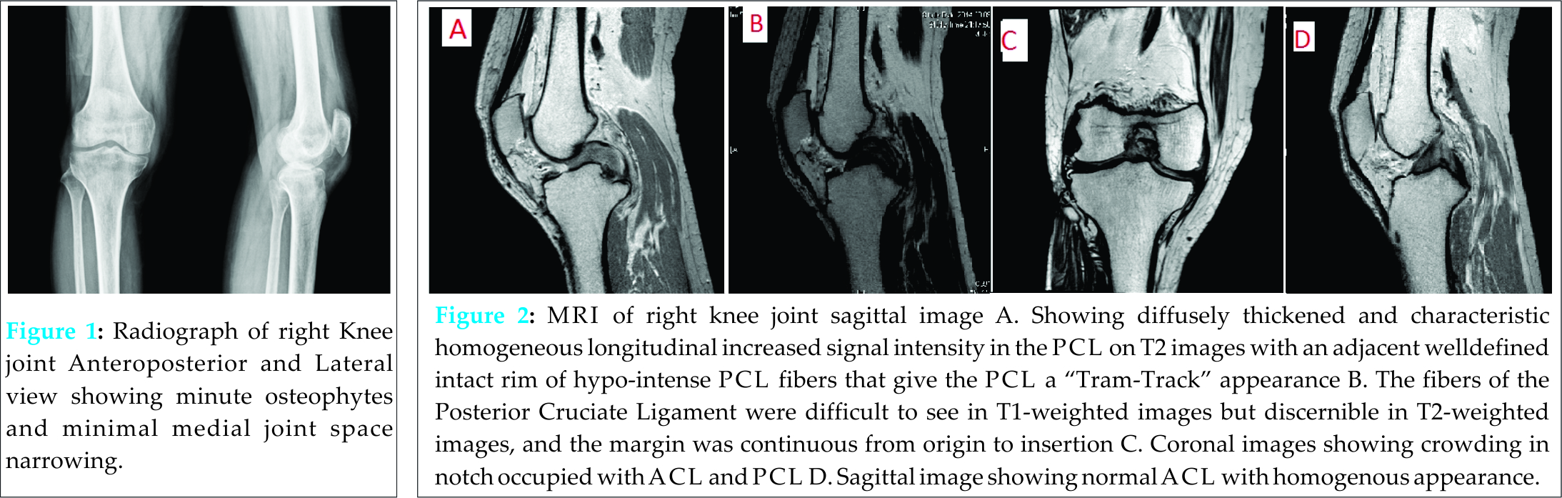
The fibers of the PCL were difficult to see in T1-weighted images but discernible in T2-weighted images, and the margin was continuous from origin to insertion. Medial meniscus was in partial menisectomy state and lateral meniscus was normal. Based on the patient’s history and the MRI findings, we suspected mucoid degeneration or an intraligamentous ganglion of the Posterior Cruciate Ligament as the cause of the patient’s symptoms.
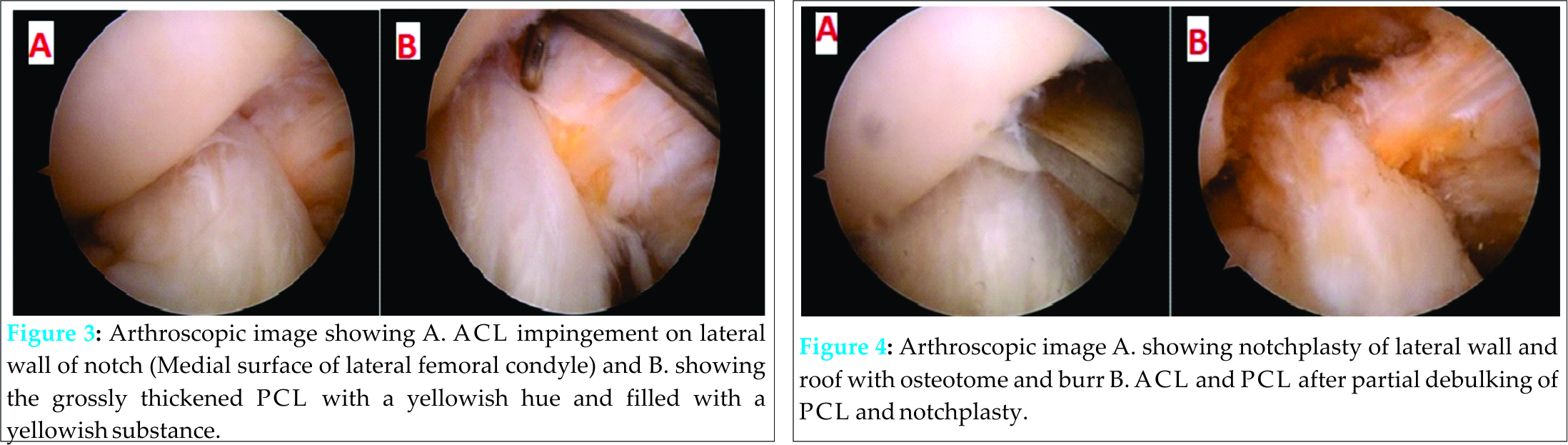
Arthroscopic examination showed PCL was grossly thickened and colored with a yellowish hue in the ligament, and was filled with a yellowish substance (Fig 3B). The yellowish hue was not liquid, but a fibrous tissue-like ACL mucoid degeneration as described in reports on ACL mucoid degeneration. Anterior cruciate ligament appearance and tension with probing was normal but was pushed towards lateral wall of the notch and was impinging on lateral wall in flexion and on roof on extension (Fig 3A). On probing, tension of the PCL fibers themselves was normal suggesting the absence of PCL tear. We carefully split the fibers and curetted the yellow tissues preserving the PCL fibers as possible (Fig 4B). We restricted curettage when the thickness of PCL decreased, because the extensive curettage would cause damage of PCL tensions . We did lateral wall and roof notchplasty and checked again for impingement in flexion and extension. After arthroscopic procedure, Lachmann and Pivot shift were negative ruling out instability. Medial meniscus was in partial menisectomy state and lateral meniscus was normal. Cartilage of femoral condyle and patella was normal. Histological examination of the biopsied tissue revealed mucoid degeneration of the ligament. Early physiotherapy rehabilitation was started from the second post-operative day. Immediately after surgery, patient was pain-free and gained full range of motion. At one year follow up, patient remained asymptomatic. Informed Consent was obtained from the patient for publication of this case report.
Discussion
Mucoid degeneration of the CLis relatively rare and therefore it is less known than its counterpart involving the ACL. It is already established from surgical and histopathological studies that degeneration of the ACL and PCL often coexists and that the former is a predictor of the latter . Mucoid degeneration of PCL has been described previously and there are few case reports but these cases are of asymptomatic patient or with PCL symptoms in terms of pain on terminal flexion . We herein report a case of Mucoid degeneration of PCL in which patient predominantly presented with ACL impingement.
Mucoid degeneration of PCL can produce symptoms of pain on flexion or restricted terminal flexion . Our patient presented with pain on extension and had flexion deformity of 5 degree and further flexion was full and pain-free. The grossly thickened Posterior PCL can push ACL towards the lateral wall and roof of notch and may lead to the secondary ACL impingement symptoms. Mechanical impingement of ACL might cause extension pain associated with limited extension .
MRI images in sagittal plane showed PCL is thickened and characteristic homogeneous longitudinal increased signal intensity in the PCL in all planes on proton density and T2 images with an adjacent well-defined intact rim of hypo-intense PCL fibers that give the PCL a tram-track appearance and on coronal images notch is crowded with hypertrophic PCL.
The arthroscopic findings of grossly thickened PCL with a yellowish hue are characteristic and the PCL was filled with a yellowish substance. The yellowish hue was not liquid, but a fibrous tissue-like Anterior Cruciate Ligament mucoid degeneration as described in reports on ACL mucoid degeneration .
We excised the yellowish substance from the PCL as precisely as possible to avoid damaging the remaining PCL fiber (Limited Debulking). We did notchplasty of lateral wall and roof to accommodate the Anterior Cruciate Ligament and avoid impingement. Partial excision of this Posterior Cruciate Ligament lesions and notchplasty resulted in immediate pain relief and improved range of motion without instability. Reduction notchplasty for hypertrophic anterior cruciate ligament mucoid degeneration leads to complete pain relief in 80% of cases while maintaining good postoperative knee stability . Histological examination of the biopsied tissue revealed mucoid degeneration of the ligament. At one year follow-up patient remains asymptomatic.
Conclusion
Mucoid degeneration although asymptomatic in majority of cases may present with pain and deformity. Posterior cruciate ligament may enlarge significantly and may push the ACL in the notch and may lead to the ACL impingement symptoms. Partial Debulking of PCL and notchplasty is effective treatment with immediate postoperative pain relief and good functional results.
Clinical Message
Mucoid degeneration of ACL and PCL is a well-known entity. Correlation between clinical, radiographic and arthroscopic findings is required in order to individualize the treatment approach.
References
1. Parkar AP, Vanhoenacker FM, and Adriaensen ME, Bilateral mucoid degeneration of the posterior cruciate ligaments. JBR-BTR 2013;96(5): p. 298-300.
2. Papadopoulou P. The celery stalk sign. Radiology 2007;245(3): 916-7.
3. Okazaki K et al., Mucoid degeneration of the posterior cruciate ligament: a case report. Knee Surg Sports Traumatol Arthrosc 2011;19(1): 105-7.
4. Shoji T, Fujimoto E, and Sasashige Y. Mucoid degeneration of the posterior cruciate ligament: a case report. Knee Surg Sports Traumatol Arthrosc 2010;18(1): 130-3.
5. Viana SL et al. Diffuse intrasubstance signal abnormalities of the posterior cruciate ligament: the counterpart of the mucoid degeneration of the anterior cruciate ligament? A case series. JBR-BTR 2008;91(6): 245-8.
6. Hodler J et al. The cruciate ligaments of the knee: correlation between MR appearance and gross and histologic findings in cadaveric specimens. AJR Am J Roentgenol 1992;159(2): 357-60.
7. Kleinbart FA et al. Histologic comparison of posterior cruciate ligaments from arthritic and age-matched knee specimens. J Arthroplasty 1996;11(6): 726-31.
8. McMonagle JS et al. Tram-track appearance of the posterior cruciate ligament (PCL): correlations with mucoid degeneration, ligamentous stability, and differentiation from PCL tears. AJR Am J Roentgenol 2013;201(2): 394-9.
9. Cho SD et al. Mucoid degeneration of both ACL and PCL. Clin Orthop Surg 2012;4(2): 167-70.
10. Shoji T, Fujimoto E, and Sasashige Y. Mucoid degeneration of the posterior cruciate ligament. Knee Surg Sports Traumatol Arthrosc 2010;18(7): 1001-2.
11. Cha JR et al. Symptomatic mucoid degeneration of the anterior cruciate ligament. Knee Surg Sports Traumatol Arthrosc 2013;21(3): 658-63.
12. Morice A et al. Reduction plasty for hypertrophic anterior cruciate ligament mucoid degeneration: clinical and knee laxity outcomes in 23 cases. Orthop Traumatol Surg Res 2013;99(6): 693-7. degeneration: clinical and knee laxity outcomes in 23 cases. Orthop Traumatol Surg Res 2013;99(6): 693-7.mucoid degeneration: clinical and knee laxity outcomes in 23 cases. Orthop Traumatol Surg Res, 2013. 99(6): p. 693-7.
| How to Cite This Article: Jangir RR, Wang JH. Mucoid degeneration of Posterior Cruciate Ligament with Secondary Impingement of Anterior Cruciate Ligament: A rare case report. Journal of Orthopaedic Case Reports 2015 Oct-Dec;5(4): 44-46. Available from: https://www.jocr.co.in/wp/2015/10/01/2250-0685-343-fulltext/ |

[Full Text HTML] [Full Text PDF] [XML]
[rate_this_page]
Dear Reader, We are very excited about New Features in JOCR. Please do let us know what you think by Clicking on the Sliding “Feedback Form” button on the <<< left of the page or sending a mail to us at editor.jocr@gmail.com


