[box type=”bio”] What to Learn from this Article?[/box]
The use of a proximally based flexor hallucis longus flap is an effective alternative surgical approach that may be useful for the repair of large soft-tissue defects in the distal third of the lower leg if insufficiently covered by other flaps.
Case Report | Volume 7 | Issue 2 | JOCR March – April 2017 | Page 70-73| Tomohiro Yasuda, Masayuki Arai, Kaoru Sato, Koji Kanzaki. DOI: 10.13107/jocr.2250-0685.756
Authors: Tomohiro Yasuda[1], Masayuki Arai[1], Kaoru Sato[1], Koji Kanzaki[1]
[1] Department of Orthopaedics, Fujigaoka Hospital, Showa University School of Medicine, Yokohama, Japan.
Address of Correspondence:
Dr. Tomohiro Yasuda,
1-30 Fujigaoka, Aoba-ku, Yokohama 227-8501, Japan, Fujigaoka Hospital,
Showa University School of Medicine, Yokohama, Japan.
E-mail: shapedbytomo@gmail.com
Abstract
Introduction: In the treatment of Gustilo Type 3B open tibial fractures, it is important to perform soft tissue reconstruction and bone reconstruction simultaneously. Gastrocnemius muscle and soleus muscle flaps are generally used as rotational flaps for the tibia. The distal third of the tibia can often not be covered with the gastrocnemius muscle and soleus muscle flaps. Treatment distal to the distal third of the tibia is difficult because fewer flap options are available. In the present report, we describe our experience with a Gustilo Type 3B open tibial fracture treated by gastrocnemius muscle and soleus muscle flaps, along with an additional proximally based flexor hallucis longus flap, which is a rare procedure.
Case Report: The participant was a 17-year-old male who injured his left tibia in a motorcycle traffic accident. Physical examination revealed a wound of 13 cm × 7 cm extending from the medial lower leg to the posterior aspect, with extensive skin loss. There was no nerve or vascular injury. The tibia was exposed, with detachment of the periosteum. The radiograph revealed a tibial shaft fracture. The AO/OTA classification was 42-A3.3, and it was classified as a Gustilo-Anderson Type 3B fracture. Gastrocnemius muscle and soleus muscle flaps were lifted in the area of the soft-tissue defect and then, placed over the tibia. Despite this, the distal portion of the tibia remained uncovered. Therefore, a flexor hallucis longus flap was lifted and placed over the distal portion of the tibia. On day 7 after the injury, the external fixation device was removed and the tibial shaft was fixated with two Ender nails (4.5 mm in diameter). The clinical course was satisfactory, and the skin graft and flap were successful. Bone union was achieved without infection, and the resulting range of motion was normal.
Conclusion: For the treatment of Gustilo-Anderson Type 3B open tibial fractures, early treatment of the soft-tissue defect is vital. We surgically treated a Gustilo-Anderson Type 3B open tibial fracture with gastrocnemius muscle and soleus muscle flaps, along with an additional proximally based flexor hallucis longus flap, which is a rare procedure. In the event of a soft-tissue defect in the distal third of the tibia, the use of a proximally based flexor hallucis longus flap is an effective surgical approach.
Keywords: Tibial fracture, open fracture, flexor hallucis longus flap, Gustilo-Anderson Type 3B facture.
Introduction
The treatment of Gustilo-Anderson Type 3B open tibial fractures is challenging for orthopedic surgeons and is associated with various difficulties. These injuries involve severe soft tissue damage and complex fractures and are associated with complications. Gustilo et al. reported that the rate of infection in Type 3B open fractures is approximately 50% [1]. When the incorrect treatment approach is selected for open tibial fracture repair, it can lead to infection, osteomyelitis, and amputation. Godina reported that in patients with open fractures, a free flap transfer within 72 h of injury results in good outcomes, with an infection rate of 1.5% [2]. Hertel et al. reported that for Gustilo-Anderson Type 3B and Type 3C open tibial fractures, superior results for both infection and bone union rates were observed in the group able to undergo immediate reconstruction within 24 h of injury compared with the group unable to undergo immediate reconstruction (mean 4.4 days) [3]. In open tibial fractures, both bone and soft tissue sustain injuries. Therefore, it is important to proceed with bone reconstruction and soft tissue reconstruction as promptly as possible. For Gustilo-Anderson Type 3B open tibial fractures, reconstruction should ideally be performed within 24 h using free flap transfer; however, some cases can be treated with a pedicle flap. Furthermore, few institutions and practitioners are able to perform free flap transfer within 24 h. If the site of the soft tissue injury is accurately assessed and treatment with a pedicle flap is possible, this would reduce the stress on the patient and practitioner. We report our experience with a case of a Gustilo-Anderson Type 3B open tibial fracture sustained by a 17-year-old male who was treated with gastrocnemius and soleus flaps as well as a proximally based flexor hallucis longus flap.
Case Report
The participant was a 17-year-old male who suffered a left tibial injury in a motorcycle traffic accident. Physical examination revealed a wound of 13 cm × 7 cm extending from the medial lower leg to the posterior aspect, with a large skin defect. The wound was partially contaminated. The tibia was exposed, with detachment of the periosteum. The gastrocnemius and soleus were distally torn. The posterior tibial artery and tibial nerve were exposed. There was no nerve or arterial injury (Fig. 1). X-ray revealed a tibial shaft fracture (Fig. 2).

This tibial fracture was graded as follows with AO/OTA classification: 42-A 3.3 and Gustilo-Anderson Type 3B. On the hannover fracture scale, the fracture scored 5 points. The ganga hospital injury severity score was six. Photographs in Fig. 1 showing (a) injury to skin and fascia: Wound with skin los over the fracture, (b) injury to bone and joints: Wound without skin loss but exposing the fracture site, (c) injury to musculotendinous and nerve units: Partial injury to unit, and (d) comorbid conditions: None.

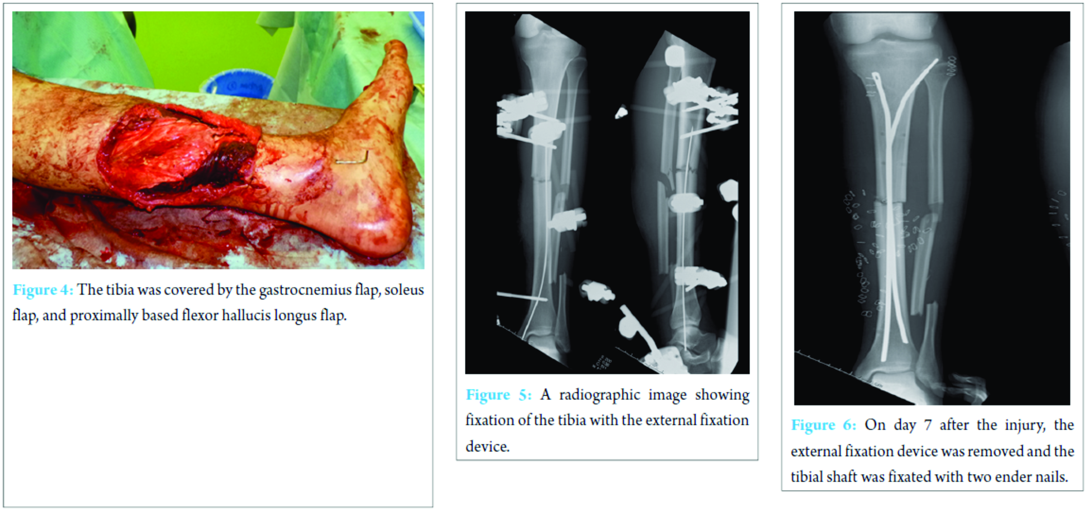
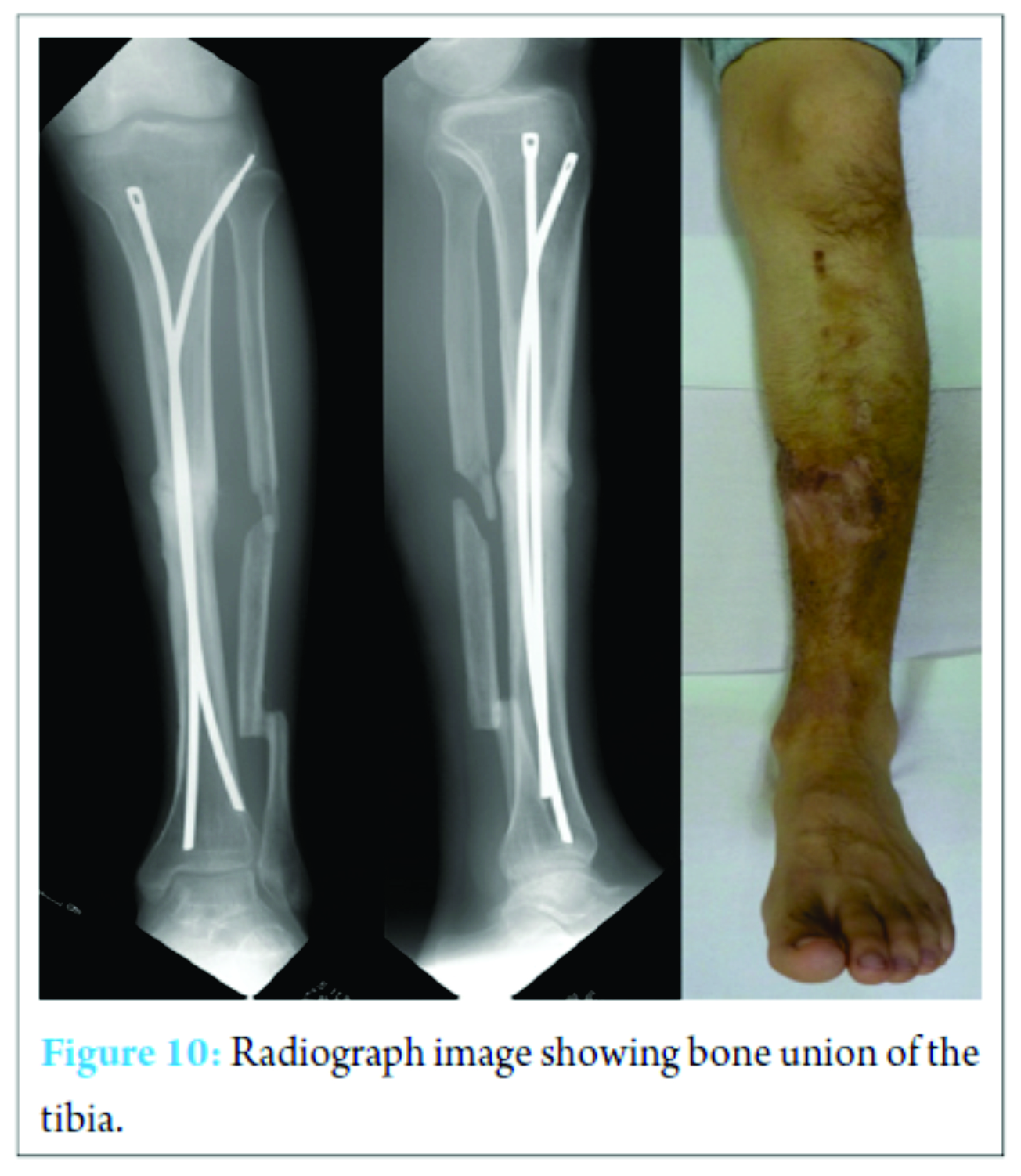
Broad-spectrum antibiotics with Gram-positive, Gram-negative, and anaerobic coverage were started intravenously. Emergency surgery was performed under general anesthesia. The patient was positioned in the supine position, and a tourniquet was used as needed. The wound was debrided and then, washed with physiological saline. The tibia was temporarily fixated using an external fixation device. We then checked that blood flow to the gastrocnemius muscle and soleus muscle was maintained. The gastrocnemius and soleus muscle flaps were lifted and placed over the tibia. Despite this, the distal portion of the tibia was insufficiently covered. Therefore, a flexor hallucis longus flap was lifted and placed over the distal portion of the tibia (Fig. 3). Through the wound, the flexor hallucis longus could be easily identified and lifted. Following the flap lifting, the flexor hallucis longus flap had adequate blood flow. The tibia was covered by the gastrocnemius and soleus flaps and the proximally based flexor hallucis longus flap (Fig. 4). The tibial fracture was fixated using an external fixation device and a 2 mm Kirschner wire (Fig. 5). At the foot, the distal portion of the flexor hallus longus was changed to the tendon of the flexor digitorum longus muscle. After 5 days, the patient was shifted to oral antibiotics. On day 7 after the injury, the wound external fixation device was removed and the tibial shaft was fixated with two Ender nails (4.5 mm in diameter). The flaps were covered with skin grafts and tie-over dressings were applied (Fig. 6 and 7). At the 2-week follow-up, the tie-over dressing was removed. At the follow-up examination at 3 weeks, the skin graft and flaps were healing successfully and rehabilitation was commenced with knee motion and gait training (Fig. 8). 1 month following surgery, compared with the healthy side, the range of movement of the hallux with the transferred tendon was almost normal (Fig. 9). At the final follow-up at 18 months, fractures were consolidated with normal range of motion. There was no angulation of more than 5°. Range of motion at the end of treatment was 35° in ankle dorsiflexion and 40° in ankle plantar flexion, 160° in knee flexion, and 0° in knee extension, symmetrical to the contralateral. The patient walked without pain and limp. The wound had healed without any infection (Fig. 10).

Discussion
Open fractures are usually associated with soft tissue injury. Moreover, severe open fractures occur as a result of high-energy trauma, such as those associated with motorcycle accidents. For adequate open fracture treatment, it is important to accurately ascertain the state of the fracture, including the assessment of the fracture, skin, muscles, nerves, and blood vessels as well as the degree of contamination. Then, a treatment plan should be created. If the fracture, skin, muscles, nerves, blood vessels, and degree of contamination can be assessed during surgery, the precise Hannover fracture scale and AO classification can be confirmed. After assessing the soft tissue defects caused by an open tibial fracture, reconstruction should be performed within 72 h [2, 4]. For soft tissue reconstruction in open tibial fractures, the type of flap used differs according to the extent of the skin defect at the fracture site. Tintle et al. and Kamath et al. created an algorithm for flap selection, in which the lower leg is divided into thirds, i.e., proximal, middle, and distal [5, 6]. Burns et al. did not observe a significant difference in the rate of infection in relation to the type of flap used, i.e., free flaps or rotational flaps, for open tibial fractures [7]. Thus, the respective advantages of and different purposes for which free flaps and rotational flaps should be used remain controversial. Skin loss in the present case extended from the middle third (middle) to the distal third (distal) of the lower leg. For such an injury, soft tissue reconstruction is generally performed with a gastrocnemius flap and soleus flap. However, the gastrocnemius flap and soleus flap do not sufficiently cover the distal area of injury (soft tissue defects at the distal third of the leg). For soft tissue defects, in the distal third of the lower leg, treatment options include a flexor digitorum longus muscle flap, distally based sural artery flap, reverse soleus flap, or distally pedicled peroneus brevis muscle flap [8, 9, 10]. Considering the zone of the injury, a flexor digitorum longus muscle flap, distally based sural artery flap, or reverse soleus flap was not suitable for our patient. When evaluating the zone of injury in open tibial fractures, the choice of flap should be made upon confirming that the blood flow is maintained in the flap. The proximally based flexor hallucis longus is usually used for reconstructing the lateral distal portion of the lower leg. In the present case, adequate blood flow was able to be confirmed through the wound; thus, we selected a proximally based flexor hallucis longus flap. A rotational flap was transferred in the initial surgery, and the wound healed without any infection. A rotational flap is easy to use because it does not require microsurgery. However, the proximity of the source of the flap and the zone of injury makes judgments difficult. As a precaution, in the event of rotational flap necrosis, a free flap should be prepared. Fujioka et al. reported that bone-exposing wounds in our patients with Gustilo-Anderson 3B and C fracture had nor improved with artificial dermis and skin grafting technique, and required conventional flap surgery. They concluded that artificial dermis is not a recommendable resurfacing option for patients with Gustilo-Anderson 3B and C fracture because the poor circulation of the bone may result in osteomyelitis [11].
Conclusion
For the treatment of open tibial fractures classified as Gustilo-Anderson Type 3B, the treatment of the soft tissue is difficult and is associated with complications. When treating bone fractures, the treatment of the soft tissue needs to proceed simultaneously. Notably, for lesions in the distal third of the tibia, few rotational flaps can be used. In general, flaps of the gastrocnemius and soleus muscles are used as rotational flaps for the lower leg. In the present case, we performed a reconstruction surgery using gastrocnemius and soleus muscle flaps and an additional proximally based flexor hallucis longus flap, which is a rare procedure. As a result, the wound healed without any infection. The use of a proximally based flexor hallucis longus flap was an effective surgical approach to treat the soft tissue defect in the distal third of the lower leg.
Clinical Message
In the treatment of open tibial fractures classified as a Gustilo-Anderson Type 3B, early treatment of the soft tissue is vital. The use of a proximally based flexor hallucis longus flap is an effective alternative surgical approach that may be useful for the repair of large soft-tissue defects in the distal third of the lower leg if insufficiently covered by other flaps.
References
1. Gustilo RB, Mendoza RM, Williams DN. Problems in the management of Type III (severe) open fractures: A new classification of Type III open fractures. J Trauma 1984;24(8):742-746.1. Gustilo RB, Mendoza RM, Williams DN. Problems in the management of Type III (severe) open fractures: A new classification of Type III open fractures. J Trauma 1984;24(8):742-746.
2. Godina M. Early microsurgical reconstruction of complex trauma of the extremities. Plast Reconstr Surg 1986;78(3):285-292.
3. Hertel R, Lambert SM, Müller S, Ballmer FT, Ganz R. On the timing of soft-tissue reconstruction for open fractures of the lower leg. Arch Orthop Trauma Surg 1999;119(1-2):7-12.
4. Gopal S, Majumder S, Batchelor AG, Knight SL, De Boer P, Smith RM. Fix and flap: The radical orthopaedic and plastic treatment of severe open fractures of the tibia. J Bone Joint Surg Br 2000;82(7):959-966.
5. Tintle SM, Gwinn DE, Andersen RC, Kumar AR. Soft tissue coverage of combat wounds. J Surg Orthop Adv 2010;19(1):29-34.
6. Kamath JB, Shetty MS, Joshua TV, Kumar A, Harshvardhan, Naik DM. Soft tissue coverage in open fractures of tibia. Indian J Orthop 2012;46(4):462-469.
7. Burns TC, Stinner DJ, Possley DR, Mack AW, Eckel TT, Potter BK, et al. Skeletal trauma research consortium (STReC). Does the zone of injury in combat-related Type III open tibia fractures preclude the use of local soft tissue coverage? J Orthop Trauma 2010;24(11):697-703.
8. Durand S, Sita-Alb L, Ang S, Masquelet AC. The flexor digitorum longus muscle flap for the reconstruction of soft-tissue defects in the distal third of the leg: Anatomic considerations and clinical applications. Ann Plast Surg 2013;71(5):595-599.
9. Karbalaeikhani A, Saied A, Heshmati A. Effectiveness of the gastrocsoleous flap for coverage of soft tissue defects in leg with emphasis on the distal third. Arch Bone J Surg 2015;3(3):193-197.
10. Ng YH, Chong KW, Tan GM, Rao M. Distally pedicled peroneus brevis muscle flap: A versatile lower leg and foot flap. Singapore Med J 2010;51(3):339-342.
11. Fujioka M, Hayashida K, Murakami C. Artificial dermis is not effective for resurfacing bone-exposing wounds of Gustilo-Anderson III fracture. J Plast Reconstr Aesthet Surg 2013;66(4):e119-e121.
 |
 |
 |
 |
| Dr. Tomohiro Yasuda | Dr. Masayuki Arai | Dr. Kaoru Sato | Dr. Koji Kanzaki |
| How to Cite This Article: Yasuda T, Arai M, Sato K, Kanzaki K. A Gustilo Type 3B Open Tibial Fracture Treated with a Proximal Flexor Hallucis Longus Flap: A Case Report. Journal of Orthopaedic Case Reports 2017 Mar-Apr;7(2):70-73. |
[Full Text HTML] [Full Text PDF] [XML]
[rate_this_page]
Dear Reader, We are very excited about New Features in JOCR. Please do let us know what you think by Clicking on the Sliding “Feedback Form” button on the <<< left of the page or sending a mail to us at editor.jocr@gmail.com


