
[box type=”bio”] What to Learn from this Article?[/box]
Presentation and management of this rare entity.
Case Report | Volume 4 | Issue 4 | JOCR Oct-Dec 2014 | Page 33-36 | Dwidmuthe SC, Nemade AS, Agrawal S, Pathak A. DOI: 10.13107/jocr.2250-0685.221
Authors: Dwidmuthe SC [1], Nemade AS [1], Agrawal S [2], Pathak A [1]
[1] Department of orthopaedics, NKP SIMS, DIgdoh Hills, Nagpur- 440019, India.
[2] Agrasen Hospital, Ganesh Nagar, Gondia, India.
Address of Correspondence:
Dr. Samir C Dwidmuthe, Department of Orthopaedics, NKP Salve IMS, Digdoh hills, Nagpur 440019, India. Email: nagpurkneeclinic@gmail.com
Abstract
Introduction: Primary synovial osteo chondromatosis (PSOC) is chondroid metaplasia with multi¬nodular proliferation of the synovial lining of a diarthrodial joint, bursa, or tendon sheath. It usually occurs in third-fifth decade and shoulder joint involvement is infrequent. It is very rare in children and primary extraarticular PSOC of the shoulder has been reported very rarely in children.
Case Report: We present a case of primary PSOC of the long head of biceps in 8year child. It presented as painful swelling in proximal arm. The pain radiograph was showing multiple calcified loose bodies on anteromedial aspect of humerus. MRI scan showed fluid filled cysts with calcified wall. The lesion was excised through deltopectoral approach. He had complete resolution of symptoms without recurrence at 1 year. The diagnosis was confirmed on histopathological examination.
Conclusion: We want to emphasize that one should keep a differential diagnosis of this rare condition in patients presenting with cystic swelling with calcified wall. We further emphasize the need to follow these patients to detect recurrence or malignant transformation.
Keywords: PSOC, Children, Biceps Tendon.
Introduction
Synovial chondromatosis (SC) is a rare condition in which cartilaginous masses are formed by metaplasia of the synovial membrane [1, 2]. It characteristically involves single joint but extra-articular forms in the form of tendon sheath or bursal [3] involvements have been reported. Secondary forms occur in which cartilage is found in the synovial membrane in association with loose bodies and degenerative joint disease. Synovial chondromatosis usually occurs in the age group of 30 yrs to 50 yrs [4] but also has been reported in children as young as 9yrs [5] and 8 years. It usually presents as swelling in or around the joint with pain and restriction of movements. Treatment is either arthroscopic or open surgical removal of the nodules and synovectomy. Franklin first reported extra articular synovial chondromatosis in 1977. SC involving shoulder joint is rare but extra articular involvement is very rare. Horti [6] has reported SC in subacromial bursa. We are reporting the case of extra articular synovial chondromatosis around shoulder joint in pediatric age group because of its rarity and therapeutic challenge.
Case Report
A healthy 8year old male child presented with a history of a gradually increasing swelling around the right shoulder joint since 2 months. It had been painful right from the beginning and associated with mild restriction of movements. He denied any history of trauma or any other systemic complaints. The swelling progressed in size over next month. On examination there was a tender swelling on the proximal anteromedial aspect of shoulder. The swelling was not fixed to underlying bone and overlying skin. The mobility of swelling was less in longitudinal direction. Abduction and forward flexion of the shoulder were painfully restricted. Radiological examination revealed multiple well defined swellings on anteromedial aspect of proximal humerus with calcified wall. One of the swelling was approximately 2cm X 2cm. This picture was suggestive of calcified hydatid cyst or chondrocalcinosis. Total blood count was 12000 with predominance of leucocytes with normal eosinophilic count. Ultrasound of abdomen and chest showed no evidence of hydatid cyst. Ultrasound guided aspiration of the large swelling was suggestive of inflammation with no evidence of infection. MRI showed well defined lesion with calcific wall with multiple loose bodies. These swelling were arising from biceps tendon sheath. MRI reported normal articular cartilage of glenohumeral articulation without any loose bodies inside the joint. Radiologist opined in favor of a synovial chondromatosis and gave a differential diagnosis of calcified hydatid cyst. There is only one reported case of extra articular synovial chondromatosis in subacromial bursa around shoulder. Considering patients symptoms we decided to excise the swelling. Under general anaesthesia the lesion was approached through delto pectoral approach. The proximal part of pectoralis major insertion was released to expose the swelling. Well defined swelling was arising from biceps tendon sheath, with multiple other small lesions. No communication was seen to shoulder joint. All the swellings were excised. To ensure complete excision intra operative image intensifier was used. Child reported complete resolution of pain at three week after the surgery. Histopathology revealed circumscribed lobulated nodules of osteocartilaginous tissue with the stroma consisting of cellular fibroblastic tissue. Some fragments were covered with synovial tissue. There was no evidence of infection or malignancy in the specimen, and this was consistent with the appearance of synovial chondromatosis. The child was symptom free at 1 year after the surgery. There was no pain and limitation of motion then.
Discussion
Synovial chondromatosis (SC), first described by Reichel1 in 1900[1] , is a rare benign disorder characterized by chondroid metaplasia with multinodular proliferation of the synovial lining of a diarthrodial joint, bursa, or tendon sheath. Large joints are more commonly affected, the knee most often followed by the hip joint. Of the 191 SC cases meta-analyzed by Bloom and Pattinson in 1951, only 10 involved the shoulder [4] .It is classified in two types: the intraarticular which is more frequent and the bodies are loose or attached to the synovium and the extraarticular or para articular type where usually one body is attached to the serous layer of a tendon sheath or bursa. Differential diagnosis includes: Synovial chondrosarcoma, chondrosarcoma, Osteochondritis dissecans, osteoarthritis, Charcot joint, Tumoral calcinosis. Milgram [7] described three phases in synovial chondromatosis. Primary SC of shoulder joint is very rare. [4]. the shoulder SC is characterized by swelling of joint with large number of cartilaginous loose bodies. Articular cartilage shows destruction. Radiology, USG and MRI helps in confirming the diagnosis. There is three case of extra articular SC in subacromial bursa reported [5, 6]. Only one case of SC in biceps tendon sheath in 10 year old boy is described in literature [9] .The peculiarity of this case was a young age of occurrence at rare site. There are various theories explaining occurrence of extra articular SC [1, 2.3] like repeated irritation, injury or some preexisting communication. . [2]. this boy did not report any injury near the right shoulder. Diagnosis is based on clinical findings and imaging studies. Radiographs show multiple calcified bodies of uniform size, evenly distributed throughout the joint, or less commonly, within bursae or tendon sheaths9. Ultrasound PSOC osteochondral nodules may be seen as hyperechoic foci with acoustic shadowing [10]. Radiographs in this boy were suggestive of synovial chondromatosis. Magnetic resonance imaging (MRI) shows typical appearance like joint effusion and intraarticular, intrabursal or a tendon sheath (tenosynovial) soft mass with internal nodules. This mass is hypo/isointense to muscle on T1- and high signal on T2- weighted images [11] .Kramer has described three subtypes based on the MR signals.MR is also useful at assessing extrinsic erosion of bone and in excluding true marrow invasion, which can be a feature of a more malign process. In our case the diagnosis was arrived with classical features of SC on magnetic resonance. The treatment options for extra articular SC are removal of loose bodies with or without removal of synovial membrane [12]. As the loose bodies arise from the synovium, some authors have reported recurrence of SC when synovial membrane is not excised [13]. Some authors have shown similar results, without recurrence, with only removal of loose bodies [12, 14] In this particular case we removed the loose bodies and did removal biceps tendon sheath. Glenohumeral joint was not opened, as the MRI was showing normal glenohumeral Synovium. Complete removal of diseased Synovium was ensured to reduce chances of recurrence. Currently literature recommends synovectomy with removal of the loose bodies in the presence of active synovitis. The recurrence rate after surgery ranges from 3.2% to 22.2%1. Radiotherapy is beneficial for recurrent lesions and inhibition of FGF-9 has been suggested as a nonoperative method of treating primary synovial chondromatosis. SC involving shoulder is treated with removal of loose bodies and synovectomy as much as possible. Some authors have reported open synovectomy with osteoarticular allograft in patients with focal erosion of articular cartilage [15]. Total shoulder arthroplasty is reserved for advanced arthritis with SC. Recurrent synovial chondromatosis at same location favors diagnosis malignant transformation to synovial chondrosarcoma [16]. The majority of synovial chondrosarcoma cases arise in pre-existing PSOC, but progression of PSOC to malignancy is considered a rare event. The largest series in the literature, reported by Davis et al. [17], is a 5%prevalence of malignant transformation. Potential differentiating features include the clinical picture, with a rapidly enlarging mass or a recurring mass following excision, true cortical destruction and periosteal reaction with permeation and infiltration into the bone marrow, metastases to the lung [17]. We need to follow this child for possible recurrence and malignant transformation.
Conclusion
PSOC involving biceps tendon although rare in children, proper clinical examination, plain radiograph and MRI would help us to diagnose this condition. Meticulous dissection and respect to anatomy would allow us complete excision of lesion with full recovery of function of joint without recurrence.
Clinical Message
We want to emphasize that one should keep a differential diagnosis of this rare condition in patients presenting with cystic swelling with calcified wall. We further emphasize the need to follow these patients to detect recurrence or malignant transformation.
References
1. Campanacci M. Bone and Soft Tissue Tumors. 2nd Edition. New York. Springer-Verlag Wien. 1999.
2. Glen Mckenzie, Nigel Raby David Ritchie. A pictorial review of primary synovial osteochondromatosis. Eur Radiol.2008; 18: 2662–2669.
3. Franklin H. Sim, David C. Dahlin, And John C. Ivins, Extra-articular synovial chondromatosis. J Bone Joint Surg, June 1977(4); Vol. 59-A, 492-495.
4. David T, Drez D J. Synovial chondromatosis of the shoulder and biceps tendon. Orthopedics. 2000; 23(6):611-613.
5. Curr JF. Synovial osteochondromatosis: two uncommon examples. BMJ. 1949; 2:1020-1022.
6. Horii M, Tamai M, Kido K, Kusuzaki K, Kubo T, Hirasawa Y. Two cases of synovial chondromatosis of the subacromial bursa. J Shoulder Elbow Surg. 2001; 10(2):186-189.
7. Milgram J W. Synovial osteochondromatosis: a histopathological study of thirty cases. J Bone Joint Surg Am.1977; Sep; 59(6):792-801.
8. Covall D J, Fowble C D. Synovial chondromatosis of the biceps tendon sheath. Orthopaedic Review. 1994; 23:902-905.
9. Murphey M D, Vidal J A, Fanburg- Smith J D, Gajewski D A. Imaging of synovial chondromatosis with radiologic-pathologic correlation. Radiographics. 2007; 27:1465–1488.
10. Campeau N G, Lewis B D. Ultrasound Appearance of Synovial Osteochondromatosis of the Shoulder. Mayo Clin Proc. 1998; 73:1079–1081.
11. Kramer J, Recht M, Deely Dm, Schweitzer M, Pathria Mn, Gentili A, Greenway G, Resnick D. MR Appearance Of Idiopathic Synovial Osteochondromatosis. J Comp Assist Tomogr. 1993; 17(5):772–776
12. Shpitzer T, Ganel A, Engelberg S. Surgery for synovial chondromatosis. 26 cases followed up for 6 years. Acta Orthop Scand. 1990; 61:567-569.
13. Ogilvie-Harris DJ, Saleh K Generalized synovial chondromatosis of the knee: a comparison of removal of the loose bodies alone with arthroscopic synovectomy. Arthroscopy. 1994 Apr; 10(2):166-70.
14. Jeffreys TE. Synovial chondromatosis. J Bone Joint Surg Br. 1967; 49:530-534.
15. Tom Trajkovsk, Ian, P. Mayne, Ben M, Deheshi, M and Ferguson. Synovial chondromatosis of the shoulder: open synovectomy and insertion of osteoarticular allograft with internal fixation to repair intraoperative glenohumeral Joint Instability. Am J Orthop. 2011; 40(8):E154-E158.
16. Anract P, Katabi M, Forest M, Benoit J, Witvoet J, Tomeno B. Synovial chondromatosis and chondrosarcoma. A study of the relationship between these two diseases. Rev Chir Orthop Reparatrice Appar Mot.1996; 82(3): 216-
17. Davis R I, Hamilton A, Biggart J D. Primary Synovial Chondromatosis: a clinicopathologic review and assessment of malignant potential. Hum Pathol.1998; Jul; 29(7):683-8.
| How to Cite This Article: Dwidmuthe SC, Nemade AS, Agrawal S, Pathak A. Rare Case of Extra-articular Synovial Chondromatosis of Biceps Tendon Sheath in 8 years Male Child – A Literature Rivew. Journal of Orthopaedic Case Reports 2014 Oct-Dec;4(4): 33-36. Available from: https://www.jocr.co.in/wp/2014/10/21/2250-0685-221-fulltext/ |
(Figure 1)|(Figure 2)|(Figure 3)|(Figure 4)|(Figure 5)|(Figure 6)
[Abstract] [Full Text HTML] [Full Text PDF] [XML]
[rate_this_page]
Dear Reader, We are very excited about New Features in JOCR. Please do let us know what you think by Clicking on the Sliding “Feedback Form” button on the <<< left of the page or sending a mail to us at editor.jocr@gmail.com
Related Articles in Journal of Orthopaedic Case Reports
 October 21, 2014 Rare Case of Extra-articular Synovial Chondromatosis of Biceps Tendon Sheath in 8 years Male Child
 July 1, 2025 A Novel Technique of Arc Fixator for Supracondylar Humerus Fractures in Older Children: A Case Series
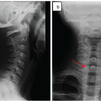 February 1, 2025 Clay-shoveler’s Fracture in a 13-year-old Child – Case Report and Review of Literature
 October 1, 2024 Brodie’s Abscess of the Humeral Paddle in a Child: A case Report


